
Figure 7.
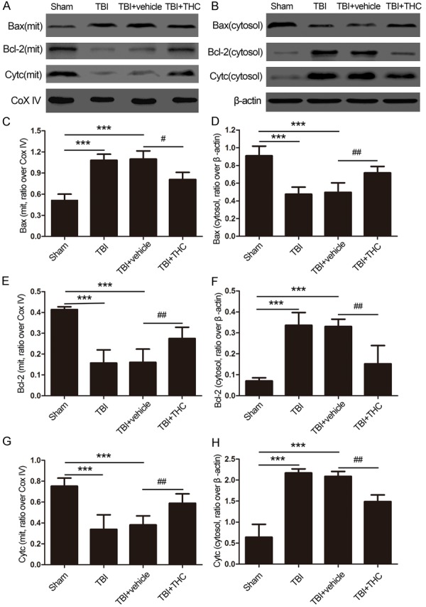
The effect of THC on mitochondrial apoptotic associated proteins at 24 h after TBI in rats. The expression of Bax, Bcl-2 and cytochrome c in mitochondria (A) and in cytoplasm (B) was evaluated by western blot analysis. Quantitative analysis the ratios of Bax (C), Bcl-2 (E) and cytochrome c (G) in mitochondria and in cytoplasm (D, F, H), normalized against Cox IV or β-actin. Data are presented as mean ± SD (n = 5 rats per group). ***P < 0.001 vs. Sham group; #P < 0.05, ##P < 0.01 vs. TBI + vehicle group.