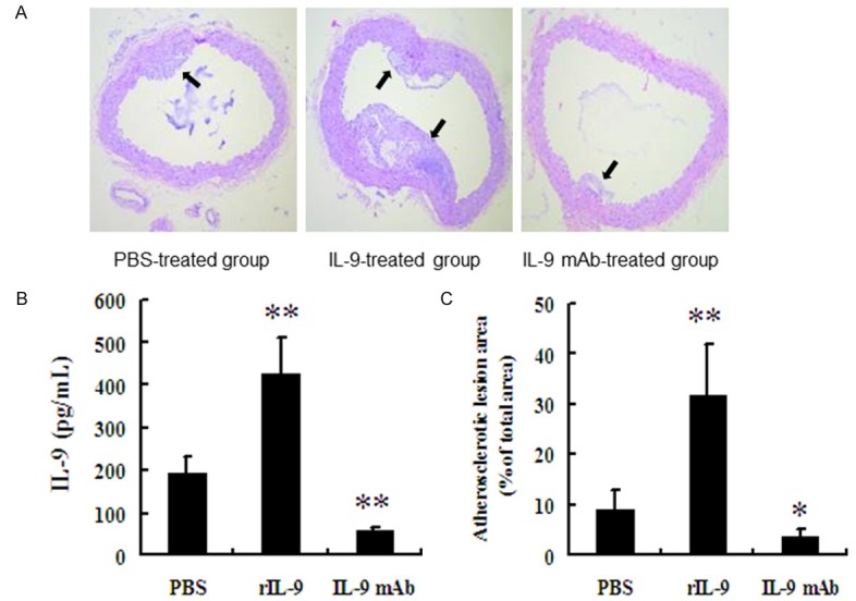
Figure 5.

Atherosclerotic lesion areas are significantly increased in mice treated with rIL-9. A. Representative micrographs of H&E staining of aorta samples (Magnification 100×). B. Quantification of aortic lesion areas as percentage of total aortic areas in various groups. C. Serum IL-9 levels in various groups. *P<0.05 vs. PBS-treatment group; **P<0.01 vs. PBS and IL-9 mAb-treatment groups.
