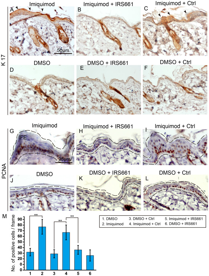
Figure 2. TLR7 activated by imiquimod induced ectopic expression of K17 and stimulated interfollicular cell proliferation.
(A–F) Immunohistochemistry staining for K17 highlighted its expression pattern in epidermis. Arrowheads indicated the ectopic expression of K17 in A and C. (G–L) Immunohistochemistry with the proliferation associated marker PCNA showed the proliferative epidermal cells. Black dashed lines outlined the epidermal region. Imiquimod treated skin (G); Imiquimod and IRS661 treated skin (H); Imiquimod and control ODN treated skin (I); DMSO treated skin (J); DMSO and IRS661 treated skin (K); DMSO and control ODN treated skin (L). IFE, interfollicular epidermis. (M) Quantification of the proliferation of interfollicular cells in G–L. **: P < 0.01, t-test.

