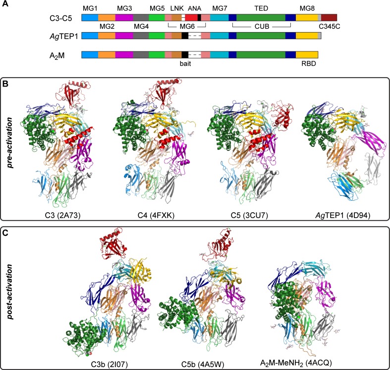
Fig. 2.
a Schematic diagram of TEP domain structure. The furin-sensitive cleavage site before the ANA domain in complement is called the ‘protease-sensitive’ region for iTEPs and the ‘bait region’ for A2M. The MG8 domain in complement and iTEPs is called the RBD for A2M. b Crystal structures of TEPs in pre-activation states: C3, C4, C5, and AgTEP1. c Crystal structures of TEPs in post-activation states: C3b, C5b, and A2M-MeNH2. Domains in (b, c) are colored according to their position in (a)

