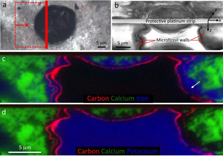
Figure 2. Mineral zonation in and around a well-preserved eukaryote microfossil from the Cailleach Head Formation.
(a) Microfossil overview showing the location of the FIB-milled area and direction of view for (b–d). (b) Secondary electron image showing the FIB-milled face below the surface of the thin section (lower half of image). (c–d) Energy-dispersive X-ray (EDS) elemental maps of the FIB-milled face shown in (b): carbon (red) represents the organic microfossil walls, highlighting a thick inner cyst wall and thinner outer vegetative cell wall; and calcium (green) represents calcium phosphate. In (c), iron (blue) represents Fe-rich clay, occurring between the two microfossil walls, replacing parts of the outer wall (arrow), and continuing for 1–2 μm outside the outer wall. In (d), potassium (blue) represents K-rich clay restricted to the interior of the vesicle. EDS spectra and electron diffraction patterns giving the complete chemistry of each of these minerals are shown in Figures 3 and 7.

