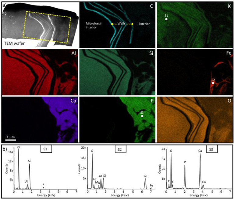
Figure 3. Mineral zonation associated with a eukaryote microfossil (Leiosphaeridia crassa) with multi-layered walls from the Cailleach Head Formation.
(a) Overview of a focused ion beam (FIB) milled TEM wafer through the microfossil wall, plus ChemiSTEM elemental maps of the boxed area. Note the multiple wall layers picked out by the carbon map. The interior of the microfossil and the area between the wall layers is filled with K-rich clay. There is also some K in the carbonaceous microfossil walls themselves and minor K-rich clay outside of the microfossil. Fe-rich clay and calcium phosphate occur in contact with the outer wall of the microfossil, with the latter being the dominant phase outside of the microfossil. (b) STEM-EDS spectra of the K-rich clay (S1), Fe-rich clay (S2) and calcium phosphate (S3) phases associated with the microfossil. Note the presence of F and C peaks in S3 indicative of francolite (carbonate fluorapatite). The positions of the spectral analyses are marked in the K, Fe and P maps respectively.

