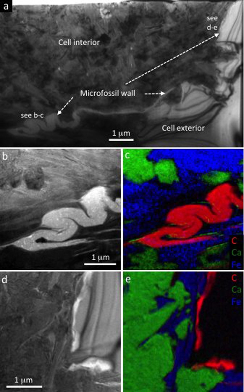
Figure 6. Nano-scale structure and chemistry of a typical prokaryote cell from the Cailleach Head Formation.
(a) Bright-field TEM image of an ultrathin section through part of a coccoid cell. The cell wall runs from top right to bottom left of the field of view, and this is folded (lower left) and fractured (right). (b, d) Bright-field TEM and (c, e) energy-filtered TEM elemental maps of two parts of the cell wall and surrounding minerals. Calcium (green) represents calcium phosphate, iron (blue) represents Fe-rich clay, and carbon (red) represents organic material of the cell wall. Both calcium phosphate and Fe-rich clay occur in direct contact with the cell wall, and are inter-grown both within and outside the cell. Quartz occurs outside the cell (black area in (e)) where the cell wall is more poorly preserved.

