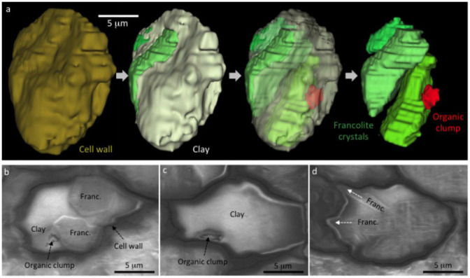
Figure 8. Microfossil preservation quality in clay minerals.

(a) 3D reconstruction of a prokaryote cell. Overview of the carbonaceous cell wall exterior is shown on the left. Next, the cell wall is removed to show the cell interior mineralized by clay (white) and three calcium phosphate (francolite) crystals (green). The clay portion is then made transparent to show that clays enclose degraded remains of putative cell contents (red). Finally (right), the clays are removed to show that the putative cell contents (organic clump) are not enclosed by francolite. (b) A single FIB-SEM slice through the microfossil reconstructed in (a), showing the 2D relationship between the cell wall, francolite, clay minerals, and the organic clump interpreted as putative cell contents. (c) A second example of a prokaryote cell with putative cell contents preserved in clay. (d) A prokaryote cell fossilized by a mixture of clay and francolite, showing how the growth of two elongate francolite crystals (arrows) caused modification of the cell wall.
