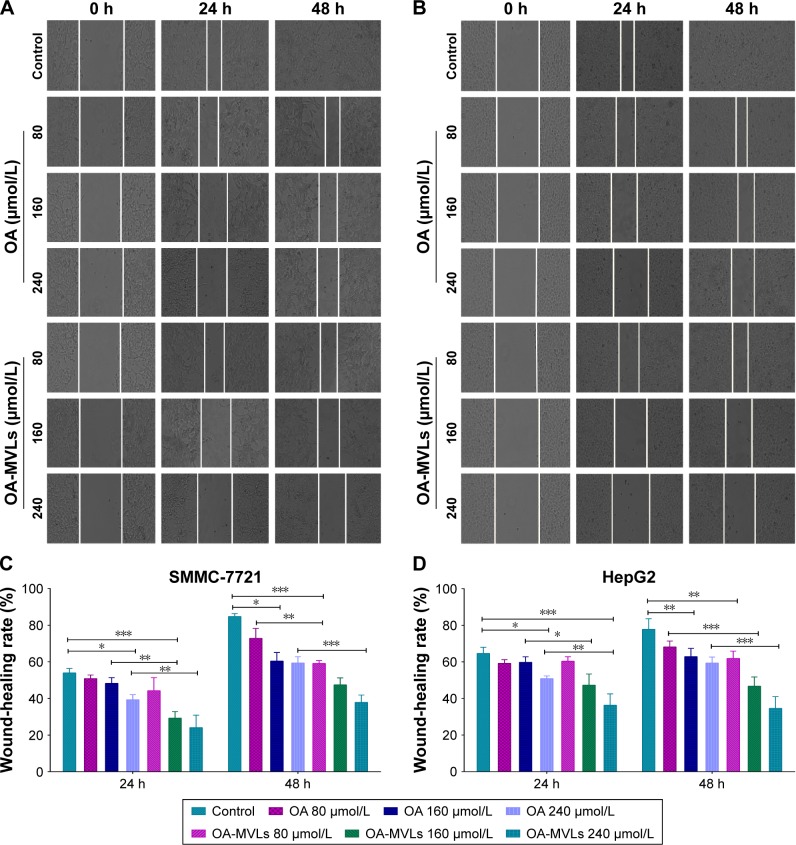
Figure 5.
Results from wound-healing assays on SMMC-7721 cells and HepG2 cells.
Notes: Cells were seeded in 6-well plates and incubated overnight. A linear area of attached cells was removed by a pipette tip before treatment with different concentrations of OA and OA-MVLs. (A) The SMMC-7721 cells and (B) HepG2 cells were photographed at different time points of 0, 24 and 48 h (100×). Meanwhile, the SW, distance between the cell fronts on either side of the wound, was measured. Wound-healing rate of (C) SMMC-7721 cells and (D) HepG2 cells was estimated according to the formula as: Wound-healing rates (%) = (SW0 h − SW24 h or 48 h)/SW0 h ×100. Each experiment was performed at least three times; results are presented as mean ± SD with *P<0.05, **P<0.01 and ***P<0.001.
Abbreviations: OA, oleanolic acid; MVLs, multivesicular liposomes; SW, scratch width; SD, standard deviation.

