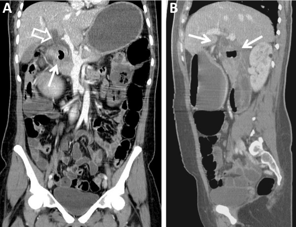
Figure 2. Contrast-enhanced Coronal and Sagittal CT of the Abdomen and Pelvis (Pre-Treatment).

Contrast-enhanced reformatted images of the abdomen and pelvis demonstrate (A) an inflamed periampullary diverticulum, which obstructs the common bile duct (arrow). The common bile duct (open arrow) is significantly dilated. In addition, intrahepatic biliary ductal dilation is seen. (B) The sagittal image provides another view of the duodenal diverticulitis and common bile duct dilation (arrows).
