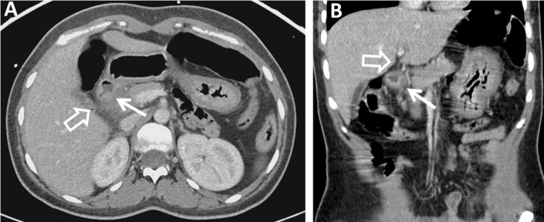
Figure 4. Contrast-enhanced Axial (A) and Coronal (B) CT Images of the Abdomen and Pelvis (Post-Treatment).

Contrast-enhanced axial (A) and coronal (B) CT images of the abdomen and pelvis two weeks after conservative treatment demonstrate a small duodenal diverticulum with significantly improved surrounding inflammatory changes (arrows). Intrahepatic biliary ductal dilation is significantly improved (open arrow).
