
Figure 2. Comparison of solution structure simulations of the TZF domains from human TTP with the family members from S. pombe and C. odorata.
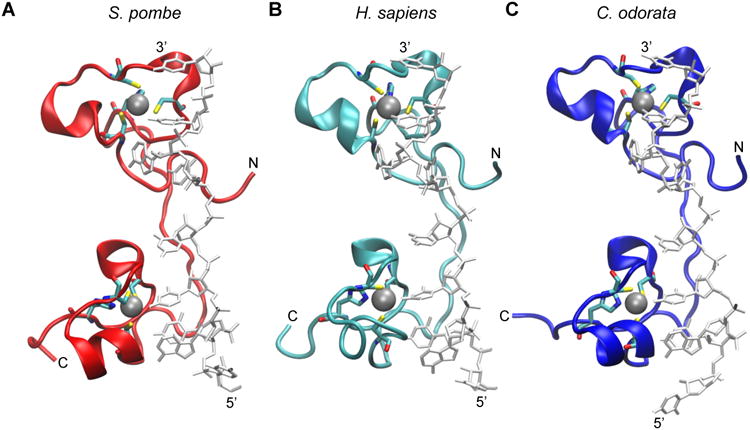
The RNA-bound structure of the TZF domain of ZFP36L2 (TIS11d) (PDB id: 1RGO) [20] was used to construct the models of RNA-bound TZF domains from: (A) S. pombe Zfs1 (GenBank accession number NP_596453); (B) human TTP (NP_003398.1); and (C) the TTP family member from C. odorata (translation of Unigene mRNA sequence GACH01022939.1), as described previously [72]. The peptide backbone of each TZF domain is represented in ribbon form, and the RNA is in stick representation in light grey. Zinc ions (silver spheres) are also shown with their coordinating residues (in stick form). Hydrogens were removed for clarity. The N and C termini of each peptide are indicated, as are the 5′ and 3′ ends of the bound RNA oligonucleotide.