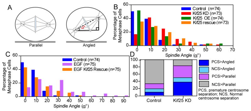
Figure 4.
Spindle orientation is altered following premature centrosome separation. (A) Schematic showing the difference between parallel and angled spindles and the measurement of spindle orientation angle, α°, relative to the coverslip. (B) Histogram of measured spindle orientation profiles of cells fixed and labeled with α- and γ-tubulin following Kif25 KD, Kif25 OE, and EGFP-Kif25 rescue following siRNA KD. (C) Histogram of measured spindle orientation profiles of cells fixed and labeled with α- and γ-tubulin following EGF treatment and EGF-treated cells overexpressing EGFP-Kif25. (D) Parsed data showing the relationship between centrosome separation state and spindle angle during mitosis. Data is initially separated based on whether cells show normal centrosome separation or premature centrosome separation then each cell is classified based on whether the mitotic spindle is parallel to the coverslip or angled. Data is from live asynchronously cycling cells transiently expressing mRFP-Pericentrin and GFP-H2B (n=26 cells, Kif25 KD; n=25 cells Control).

