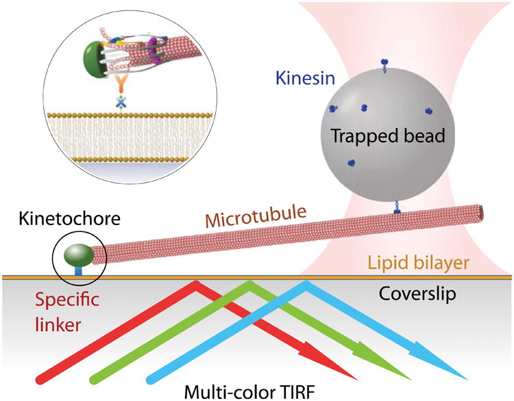
Figure 1.

Schematic of the kinetochore stretching experiment. The glass surface is passivated by a supported lipid bilayer to provide specific binding of kinetochores. A small portion of the lipid is biotinylated, which allows avidin to bind, followed by biotinylated anti-His antibody. The kinetochores are His-tagged and are therefore diffusively anchored on the surface. Microtubules spontaneously form end-on attachments to the kinetochores, and a polystyrene bead that is functionalized by kinesin is trapped optically and then bound to the free end of the microtubule. The kinetochore is dragged along the lipid bilayer by the trapped bead, and time-lapse fluorescence images of the differentially labeled kinetochore are recorded.
