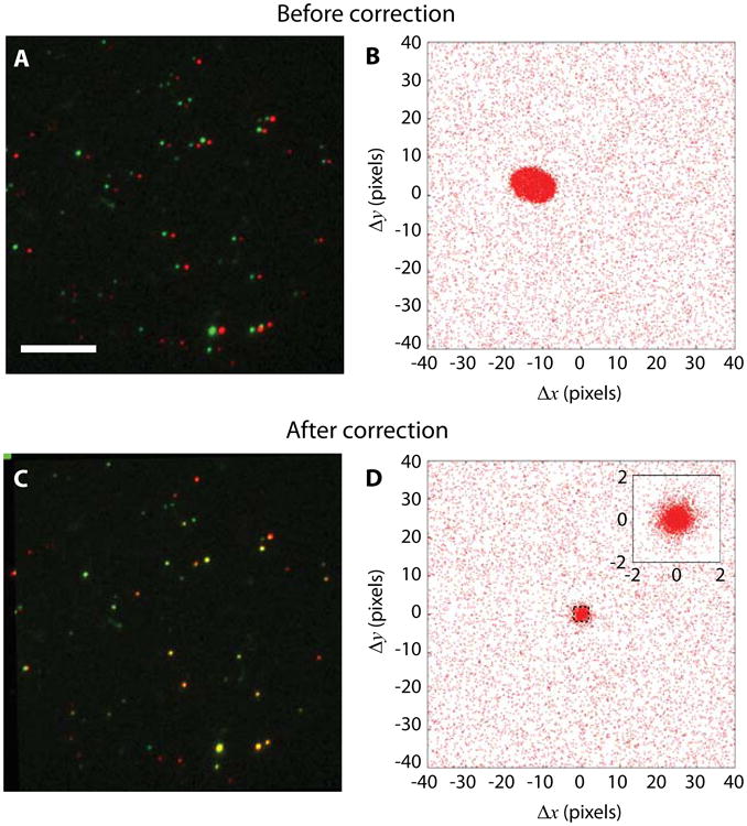
Figure 3.

Color channel position registration can be obtained directly from kinetochore imaging data without need for a separate registration step. (A) Raw images of fluorescently labelled kinetochores with Alexa Fluor 546 (false-colored in green) and Alexa Fluor 647 (false-colored in red) on two kinetochore components. Scale bar, 5 μm. (B) Pair-wise distances between all detected spots in two-color channels before registration correction. A distinctive cluster is formed near the origin. (C) Kinetochores after registration correction appear overlapping. (D) Pair-wise relative distances form a tighter cluster at the origin after correction. The inset shows the zoomed-in distribution.
