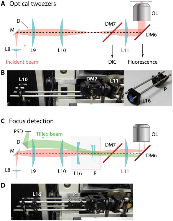
Figure 5.

Focus lock design. (A) Diagram of the optical tweezers configuration. The incident 1064 nm beam is collimated at the back-focal point of the objective lens and focused on the image plane. (B) Photograph of the actual optical elements. The extra elements for converting to a focus lock are removed, and pictured separately at right. (C) Optical diagram of the focus detection configuration. An added convex lens L16 converts the beam collimation so that the laser is focused at the back-focal plane. A wedge glass pair P steers the incident angle beyond the critical angle. (D) Photograph showing the actual parts with focus detection components inserted.
