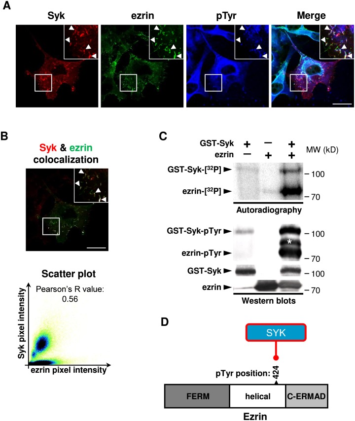
Fig 6. Syk phosphorylates ezrin on the Tyr424 residue.
(A) Syk and ezrin are both localized in pTyr-enriched plasma membrane ruffles of MDA-MB-231 cells (arrowheads). Scale bar 5 μm. (B) Quantitative analysis of Syk and ezrin colocalization with the merged picture of Syk and ezrin channels in which colocalized pixels are displayed in white (upper panel, scale bar 5 μm) and the scatter plot of pixel intensities in Syk and ezrin channels (lower panel). (C) Direct in vitro tyrosine phosphorylation of ezrin by Syk. The phosphorylation of purified Syk and ezrin (left) are analyzed by autoradiography or Western blot (pTyr). Molecular weight standards (MW) are indicated (right). *, non-specific band. (D) Cartoon illustrating the ezrin phosphorylation on Tyr424 residue by Syk. The structure of ezrin comprises a NH2-terminal FERM domain followed by a helical domain and the C-terminal actin-binding domain (C-ERMAD).

