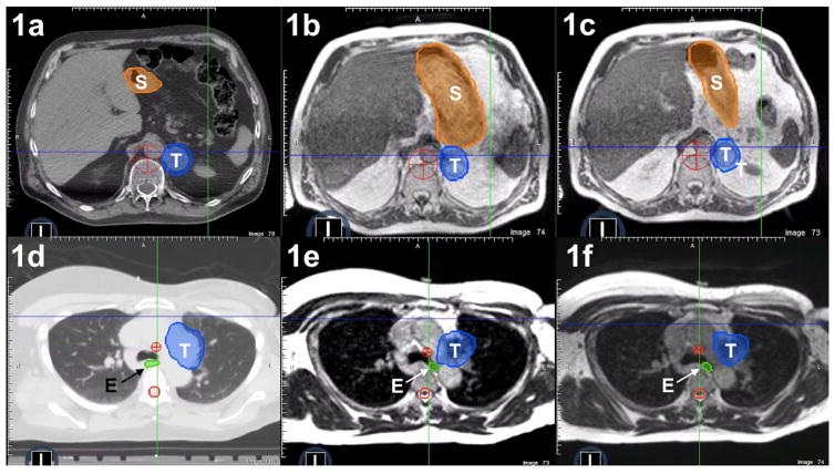
Figure 1.
Patient anatomy at simulation (1a, 1d) compared with “anatomy-of-the-day” revealed by the MR-image set at fraction one (1b, 1e) versus “anatomy-of-the-day” revealed by MR imaging at fraction two (1c, 1f). In frames 1a–1c, large interfractional shifts occur in the position of the stomach (S) relative to the adrenal tumor (T). Similarly, in a separate patient (frames 1d to 1f), interfractional variability in esophageal position (E) relative to a paraaortic lymph node tumor (T) was observed.

