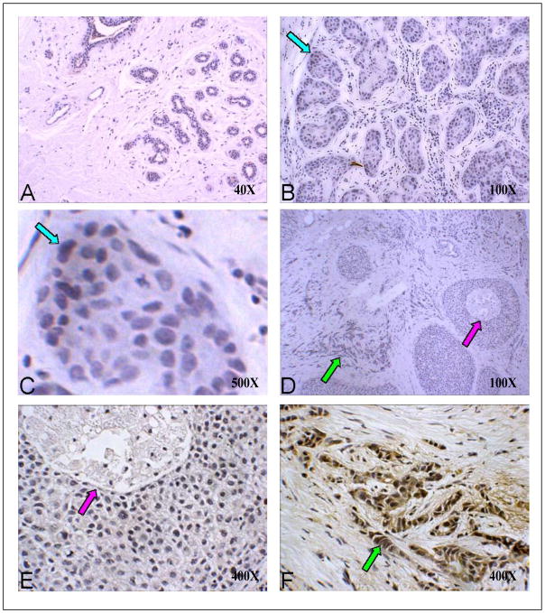
Figure 4. D11 scfv staining on normal, hyperplasia and malignant breast tissue.
A) Normal breast tissue at low (40X) magnification; B) Breast hyperplasia (100X); C) Benign hyperplasia at high (1,000X) magnification (blue arrows in B and C point to the same site at different magnifications); D) In situ and invasive breast cancer at low (40X) magnification; E) In situ carcinoma at 400X magnification (purple arrows in D and E point to the same site at different magnifications). F) Invasive cancer at 400X magnification (green arrows in D and F point to the same site at different magnifications). Note: only the invasive carcinoma in Panel F shows strong D11 scfv (brown) staining.

