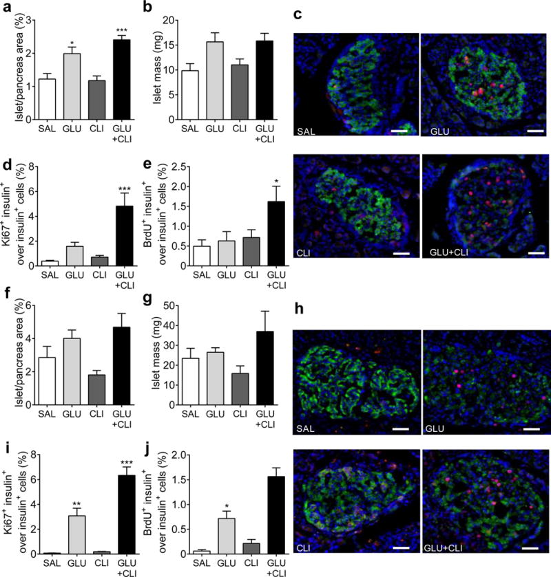
Fig. 2.

Measurement of beta cell expansion. (a–e) Two-month-old and (f–j) 6-month-old rats were infused with nutrients as indicated. (a,f) Islet area (as a percentage of total pancreas area). (b,g) islet mass. (c,h) Representative immunostaining for insulin (green), Ki67 (red) and nuclei (blue) in pancreatic sections is shown. (d,i) The percentage of Ki67+ insulin+ cells over insulin+ cells. (e,j) percentage of BrdU+ insulin+ cells over insulin+ cells was determined. Data are means±SEM (n=3–6). Scale bars, 50 μm. *p<0.05, **p<0.01 and ***p<0.001 vs SAL, ANOVA
