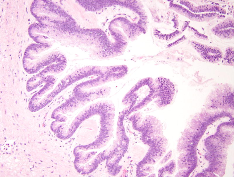
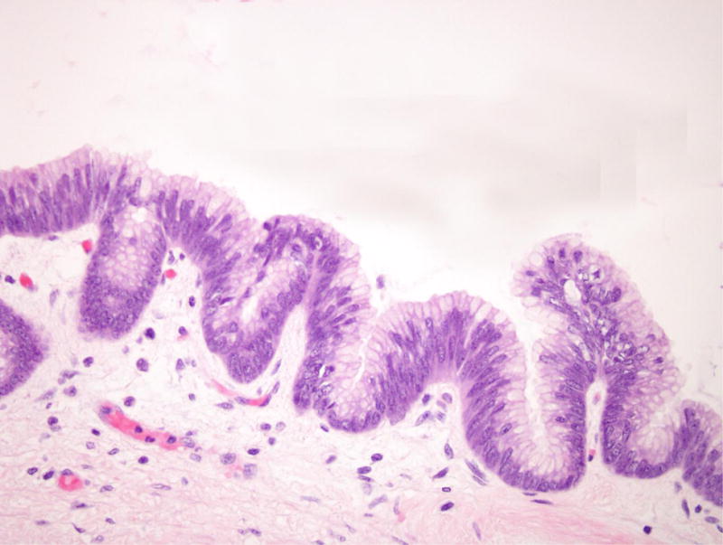
Figure 4.


Intraductal papillary mucinous neoplasm in patient 5. (A) This cystic lesion was located in the pancreatic head and shows prominent papillae. (B) The epithelial lining shows intestinal differentiation and focal high-grade dysplasia, with elongated pencillate nuclei and areas with complete loss of nuclear polarity.
