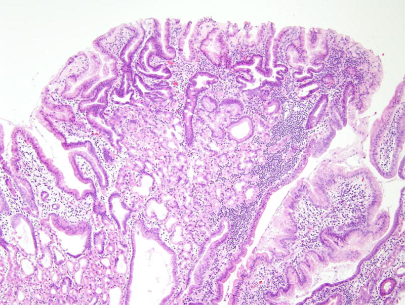
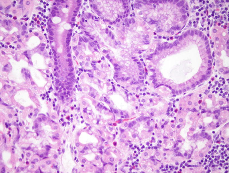
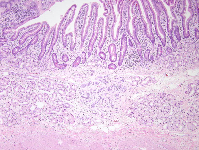
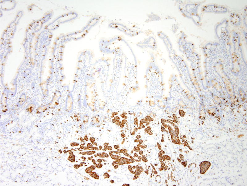
Figure 5.


Nodular gastric heterotopia and neuroendocrine proliferations in patient 5. (A) At the luminal surface, nodular gastric heterotopia in the duodenum show nodules of gastric-type epithelium. (B) No dysplasia is evident in the gastric-type epithelium. The presence of parietal cells suggests gastric heterotopia rather than gastric metaplasia. (C) Several areas of nodular gastric heterotopia had nearby underlying neuroendocrine cell proliferations. It is not clear whether these represent hyperplasia or neoplasia. (D) Synaptophysin stain confirms neuroendocrine differentiation of these cells. The largest neuroendocrine proliferation measures 10 mm.
