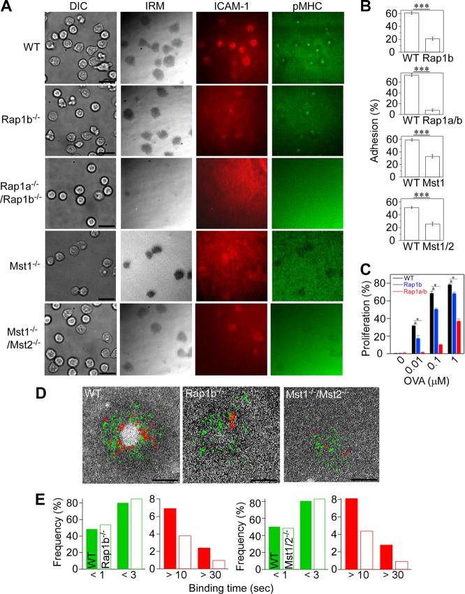
FIG 2.
Impaired IS formation in naive OT-II T cells deficient for Rap1 and Mst1/Mst2. (A) IS formation of naive OT-II T cells isolated from mutant mice deficient for Rap1b, Rap1a/Rap1b, Mst1, and Mst1/Mst2. Scale bar, 10 μm. TIRF microscopy was used to image IS formation on lipid bilayers displaying pMHC and ICAM-1. (B) Attachment of naive wild-type T cells (WT) and naive T cells from Rap1b−/− (Rap1b), Rap1a−/−/Rap1b−/− (Rap1a/b), Mst1−/−, and Mst1−/−/Mst2−/− (Mst1/2) mice. Percentages of attached cells with IRM images larger than 10 μm2 from multiple fields were counted. ***, P < 0.0001. (C) Antigen-dependent proliferation of wild-type and mutant OT-II T cells. Fractions of proliferating cells were determined by CFSE dilution after coculture with bone marrow-derived dendritic cells in the presence of OVA323–339 peptides as indicated. *, P < 0.01. (D) Single-molecule trajectories of ICAM-1 overlaid on cSMAC. Trajectories with lifetimes longer than 0.5 s (green) and 10 s (red) are shown. Scale bar, 2.5 μm. (E) Profiles of ICAM-1 binding in wild-type and Rap1b−/− or Mst1−/−/Mst2−/− OT-II cells. Short binding and long binding events are colored in green and red, respectively. The two-way chi-square P value using the Rap1b−/− or Mst1−/−/Mst2−/− data sets was <0.0001 or 0.0132, respectively.

