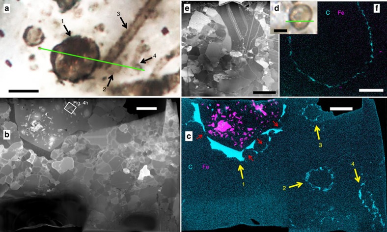
Figure 1. Iron in Gunflint microfossils.
(a–c) 1: thick-walled Huroniospora, 2: thin-walled Huroniospora, 3–4: Type 1 (cell-free sheaths) G. minuta. (d–f) Thin-walled Huroniospora. (a,d) Multiplane photomicrographs. Scale bars, 5 μm. (b,e) STEM dark-field images of the FIB ultrathin sections cut along the green lines. Scale bars, 2 μm. (c,f) STEM maps of Fe (pink), C (cyan); corresponding maps of Si, O and S are shown in Supplementary Fig. 4. Scale bars, 2 μm. Fe minerals are highly concentrated inside thick-walled Huroniospora and nearly absent in or near Type 1 G. minuta and thin-walled Huroniospora. Red arrows in c indicate displacement of wall organic matter by quartz grains.

