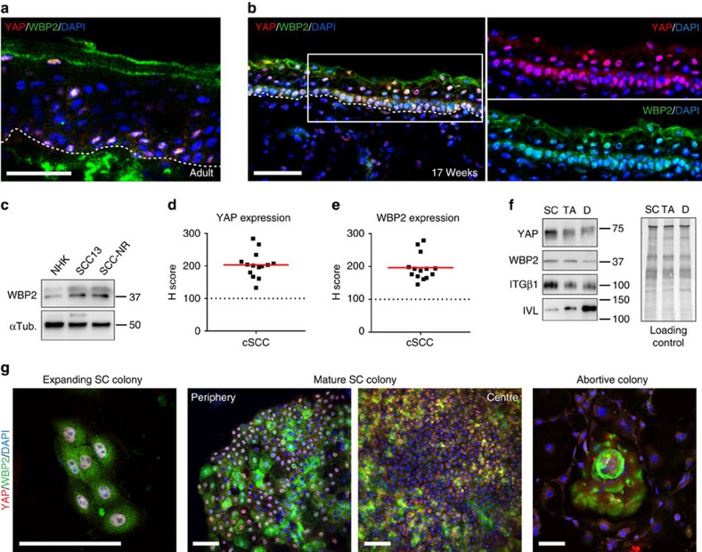
Figure 3. YAP and WBP2 are highly expressed in human epidermal SCs and upregulated in cSCC.
(a,b) Representative images (single optical planes) of adult (a) and fetal (17 weeks gestation) (b) human skin sections immunolabelled with the indicated antibodies and counterstained with DAPI to reveal nuclei. White dotted lines demarcate dermal-epidermal boundaries. Scale bars, 50 μm. (c) Western blot analysis of NHKs, SCC13 cells and primary cSCC cells (SCC-NR) using antibodies against WBP2. Tubulin was used as loading control. (d,e) Semiquantitative analysis (H-score) of YAP (d) and WBP2 (e) immunostaining intensities (individual data points) in a panel of cSCC- compared with normal skin sections (dotted line). Red lines represent the mean. (f) Western blot analysis of enriched populations of stem- (SC), transit amplifying- (TA), and terminally differentiated (D) cells using antibodies against YAP, WBP2, ITGβ1, and involucrin (IVL). Equal protein loading was confirmed by enhanced tryptophan fluorescence imaging (Bio-Rad) of PVDF membranes (loading control). (g) Representative images (maximum intensity projections) of expanding and mature SC colonies as well as abortive colonies, immunolabelled with indicated antibodies and counterstained for DAPI to reveal nuclei. Scale bars, 100 μm. PVDF, polyvinylidene difluoride.

