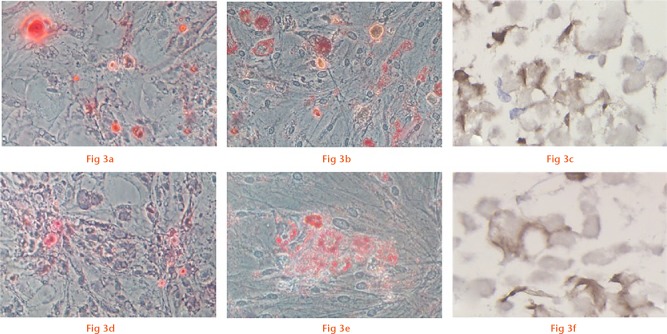
Multilineage differentiation potential of P3 cells that proliferated in normal medium (a to c) and P4 cells that proliferated in medium with low-dose G-CSF (d to f). Osteogenesis is detected by the deposition of calcium mineralisation using alizarin red staining (a, d, ×200). Adipogenesis is indicated by the accumulation of lipid vacuoles using oil red O staining (b, e, ×200). Chondrogenesis is indicated by the cartilage-specific matrix using collagen type II staining (c, f, ×400).

An official website of the United States government
Here's how you know
Official websites use .gov
A
.gov website belongs to an official
government organization in the United States.
Secure .gov websites use HTTPS
A lock (
) or https:// means you've safely
connected to the .gov website. Share sensitive
information only on official, secure websites.
