Fig. 1.

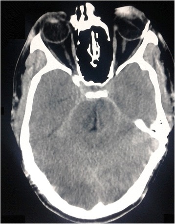
Computed tomography image of the head; the axial section of the brain is shown. The image shows linear hyperdense areas in the ambient and suprasellar cisterns suggestive of subarachnoid bleeding

Computed tomography image of the head; the axial section of the brain is shown. The image shows linear hyperdense areas in the ambient and suprasellar cisterns suggestive of subarachnoid bleeding