
Fig. 2.
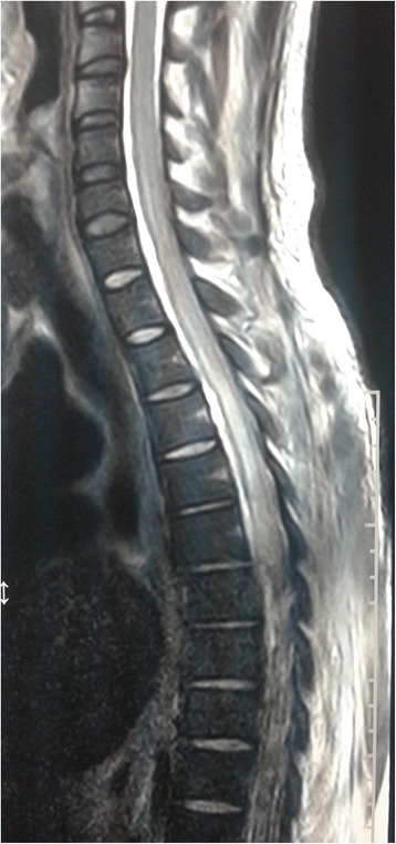
Magnetic resonance image of thoracic spine; sagittal view of thoracic segment of the spinal cord is shown. The image shows diffuse T2 high signal intensity within the spinal cord extending from second to 12th thoracic vertebral level
Official websites use .gov
A
.gov website belongs to an official
government organization in the United States.
Secure .gov websites use HTTPS
A lock (
) or https:// means you've safely
connected to the .gov website. Share sensitive
information only on official, secure websites.
Magnetic resonance image of thoracic spine; sagittal view of thoracic segment of the spinal cord is shown. The image shows diffuse T2 high signal intensity within the spinal cord extending from second to 12th thoracic vertebral level