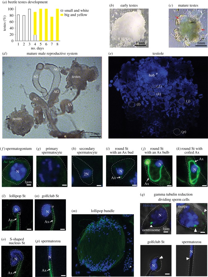
Figure 1.
Tribolium spermiogenesis. (a–c) As the beetles eclose from the pupa, their testes change (graph in a). They start as small white testes, surrounded by a big white cloud of fat (b). Around day 4, they change to large and yellow with no more than six clear lobes (testioles marked with red arrows) and very little fat (c). Though the early testes (b) appears equal in size to day 4 testes (c), in reality, the early testes fat fills the volume difference between the smaller testes and the larger ones on day 4. (d) The complete male reproductive system. (e) A single testiole stained with Hoechst (blue) to recognize the nuclei. Specific areas are presented in panels (f)–(p) as labelled. The Hoechst staining becomes lighter in later-stage sperm. (f–p) Sperm cells at distinct stages are stained with Hoechst (blue) and anti-acetylated-tubulin (green). (q) Anti-γ-tubulin (green) staining is observed in dividing-stage sperm but is undetectable in late-stage sperm (golfclub spermatid and spermatozoa) at the expected site of the centriole (arrowhead). Abbreviations: AG, accessory gland; SV, seminal vesicle; Ax, axoneme; N, nucleus; B, bulb; St, spermatid. Scale bars, 1 µm.

