Figure 3.
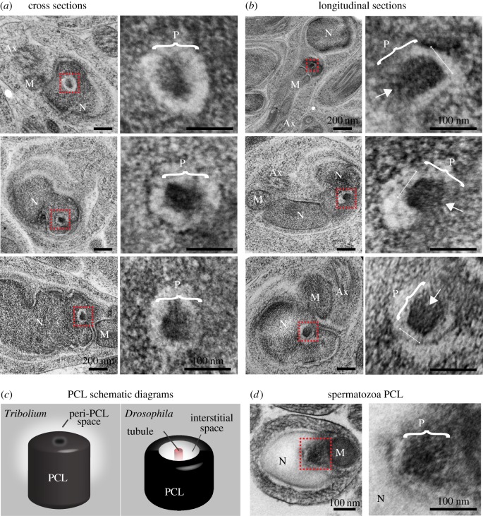
The 2nd centriole of Tribolium spermatids lack microtubules. (a,b) Representative HPF-FS TEM semi cross-section (a) and semi-longitudinal (b) analysis of Tribolium golfclub spermatids. Images on the right are insets of images on the left, signified by the red box. Brackets indicate the axis representing the PCL diameter. Arrows indicate the location of the barely detectable electron-dense central core. (c) A model of the second centriolar structure in spermatids. The structure of the PCL (P) is slightly different than Drosophila's PCL. (d) Representative TEM sections of the second centriolar structure in Tribolium spermatozoa. Abbreviations: Ax, axoneme; M, mitochondria; N, nucleus. Scale bars, 1 µm.

