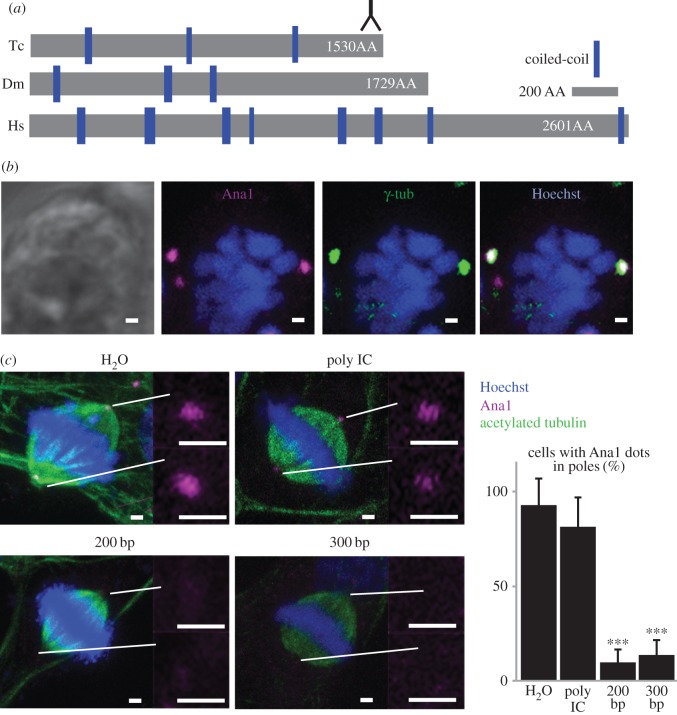
Figure 4.
Tribolium Ana1. (a) Schematic comparison of Drosophila (Dm) Ana1, Tribolium (Tc) Ana1 and human CEP295 (Hs). Inversed ‘Y’ symbol indicates the region recognized by the antibody. (b) Colocalization of γ-tubulin and Ana1 in dividing TcA cells. (c) Ana1 labelling is reduced in TcA cells when treated with two different lengths of RNAi fragments against Ana1 (200 and 300 bp), but is not diminished significantly when treated with water (H2O) or a non-specific double-stranded RNAi (Poly IC). ***p < 0.001 by t-test. n = 3. Scale bars, 1 µm.

