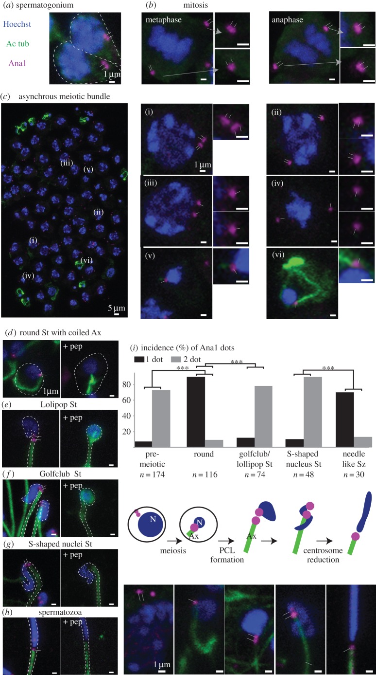
Figure 5.
A 2nd centriole forms during Tribolium spermatogenesis. (a–h) Anti-Ana1 labelling throughout Tribolium spermatogenesis. (c) As expected, an asynchronous meiotic bundle has spermatocytes, each with two centrosomes with a total of four centrioles (i–iii). Each secondary spermatocyte has one centriole at opposite poles (iv), and early spermatid stages have one centriole (v–vi). (d–h) Anti-Ana1 dots are found at the base of the nucleus throughout spermiogenesis and this staining is abolished in the presence of a competitive peptide. (i) Quantification of Ana1 dots in different stages of sperm: one dot (black); two dots (grey). A schematic diagram showing the emergence of the second centriolar structure. p < 0.001 by chi-square test. Abbreviations: Ax, axoneme; St, spermatid; Sz, spermatozoa; N, nucleus. Thin lines indicate position of centriole and PCL. Scale bars, 1 µm.

