Figure 4.

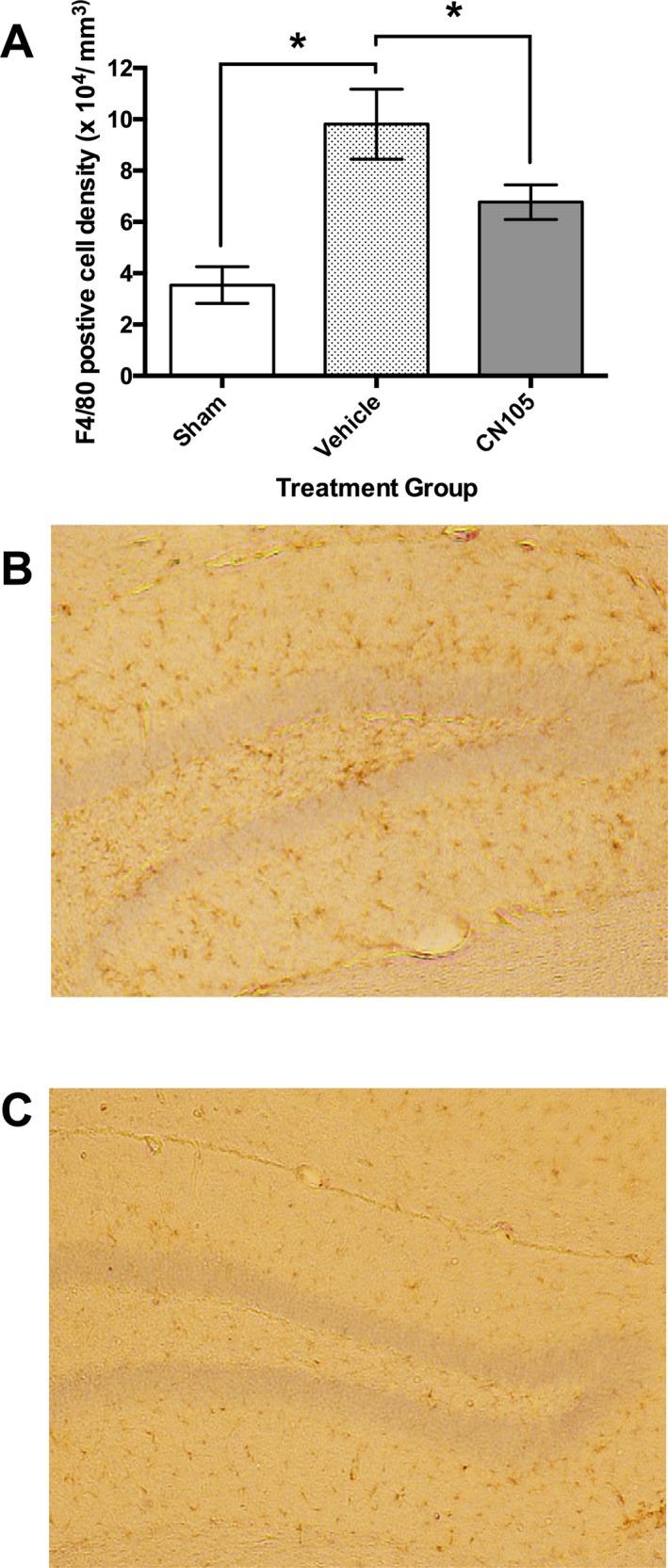
F4/80 immunocytochemical staining of microglia in contralateral hippocampus 7 days after ischemic stroke. (A) Microglial count density was significantly reduced by CN‐105, when administered 30 min post reperfusion, as compared to vehicle. Numbers indicate the number of mice that survived to 7 days in each experimental group. Representative images (4× magnification) of contralateral hippocampus demonstrating reduction in microglial cell density in hippocampal sections treated with vehicle (B) and CN‐105 (C). Microglial cells are stained brown by anti‐rat F4/80 antibody within the hippocampus. (*P < 0.05 by two‐tailed independent t‐test and error bars represent standard error of mean).
