
Figure 3. FMT ameliorates GI tract function and epithelial integrity after irradiation.
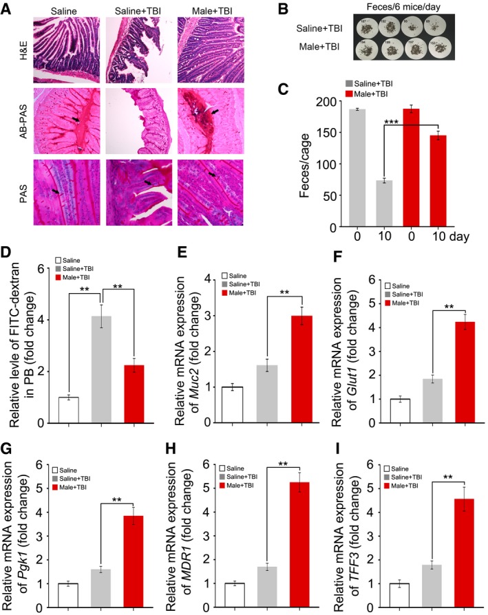
Male mice were separated into two groups after 6.5 Gy gamma ray exposure, where one cohort was treated with saline as a control and the other was treated with sex‐matched FMT.
-
AThe morphology of the small intestine in the radiation‐induced mice treated with saline or sex‐matched FMT was shown by H&E, AB‐PAS and PAS staining; the small intestine tissues were obtained at day 21 after TBI. The arrows point to the mucus layer or goblet cells.
-
B, CFaecal pellet counts removed from cage bedding on 3 day from representative cages is shown. Mean ± SD, n = 6 mice per treatment, ***P < 0.001 by Student's t‐test between Saline + TBI and Male + TBI group.
-
DThe FITC–dextran in peripheral blood from saline‐treated and sex‐matched FMT mice was assessed at day 21 after irradiation exposure. Mean ± SD. Significant differences are indicated: **P < 0.01; Student's t‐test, n = 6 per group.
-
E–IThe expression levels of Muc2, Glut1, Pgk1, MDR1 and TFF3 were examined in small intestine tissues from saline‐treated and sex‐matched FMT mice by quantitative PCR; the small intestine tissues were obtained at day 21 after TBI. Mean ± SD. Significant differences are indicated: **P < 0.01; Student's t‐test, n = 12 per group.