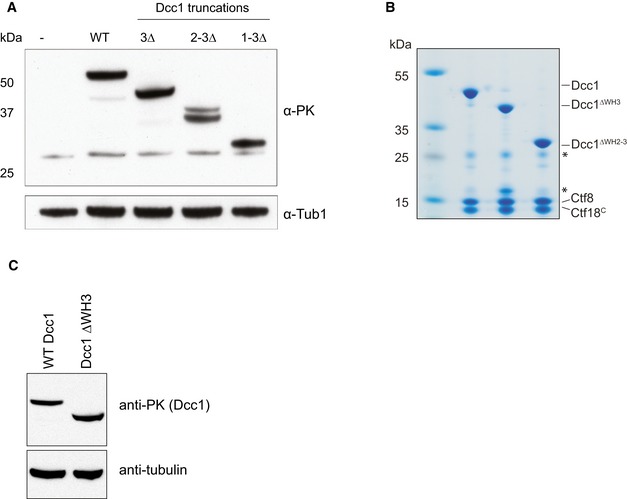
Figure EV4. Expression of and complex formation by Dcc1 WH deletions.

- Western blot showing cellular expression levels of PK‐tagged Dcc1 deletions employed for checkpoint activation and sister chromatid cohesion assays. Tubulin is shown as a loading control.
- SDS–PAGE gel of purified recombinant Dcc1‐Ctf8‐Ctf18C complexes containing indicated Dcc1 deletions. Protein complexes shown were purified in the same way as samples for crystallisation studies. Asterisks indicate impurities in the sample.
- Western blot showing cellular expression levels of Dcc1 deletion employed for ChIP analysis.
