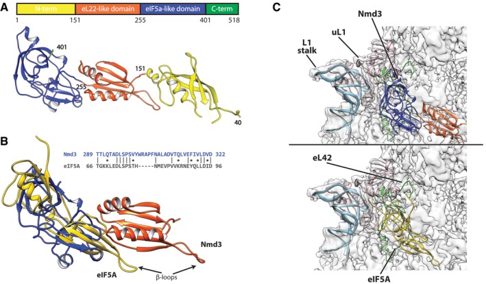
Figure 3. The structure of Nmd3.

- Linear map of Nmd3 and atomic structure colored on proposed domains. Amino acid positions are given by numbers.
- Sequence and structural alignment of eIF5a (golden) with eIF5a and eL22‐like domains of Nmd3.
- Comparison of Nmd3 from the 60S‐Nmd3‐Lsg1 complex (upper panel, colored as in A) and eIF5A (PDB: 5gak) (lower panel, golden) bound to the 60S subunit. The L1 stalk (light blue), uL1 (pink), and eL42 (green) are highlighted.
