
Figure 6. Model for coordination of Nmd3 and Sdo1 binding.
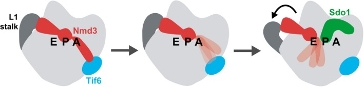
In the fully closed L1 stalk position (left), the eIF5A domain of Nmd3 is engaged with the E site, while the eL22‐like domain occupies the P site. This position is stabilized by the interaction between the N‐terminus of Nmd3 with Tif6. We propose that breaking the linkage between Nmd3 and Tif6 destabilizes the N‐terminus of Nmd3 (middle), allowing the L1 stalk to retract. Nmd3 is moved out of the P site in the retracted position, allowing Sdo1 to bind (right).