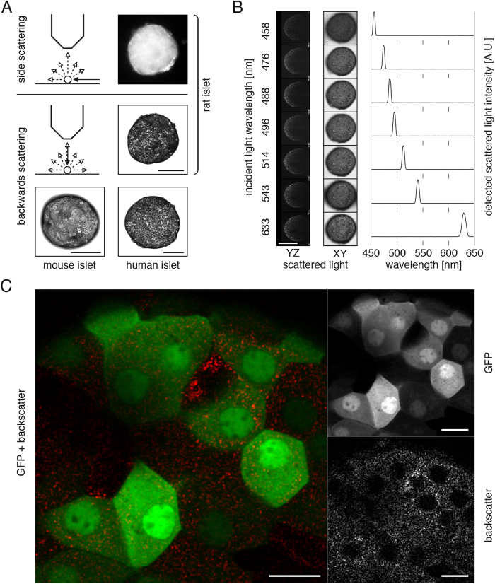
Figure 1. Pancreatic islets can be visualized using their intrinsic backscattering properties.
(A) Intrinsic scattering properties of pancreatic islets from various species allow their visualization in vitro by imaging side scattering of light by optical microscopy and backwards scattering of a laser beam scanning by confocal microscopy. This latter imaging technique not only offers high-resolution imaging but also provides three-dimensional information by the imaging of optical slices. (B) Different laser illumination wavelengths can be used to acquire backscatter images, the maximum signal intensity being collected at the incident light wavelength. (C) Confocal imaging of MIP-GFP islets at higher magnification reveals a punctuated pattern of backscatter signal, excluded from cell nuclei. The backscatter signal is present non-exclusively in each GFP-positive cell. Confocal images are presented as maximum intensity projections (A, B) or single optical planes (C). Size bars = 100 μm (A, B), 10 μm (C).

