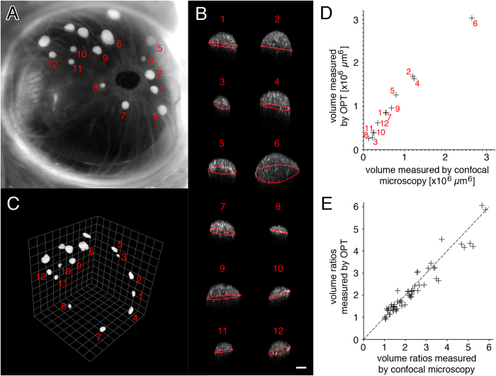
Figure 2. Backscatter signal can be used as an intrinsic indicator for islet volume.
(A) Photography of a transplanted eye shows a number of individual islets of various dimensions. (B) The islets were analyzed individually by computational analysis after in vivo imaging (islets represented as maximum intensity projection on XZ; white is backscatter signal; red line is equatorial area). (C) Mice were then sacrificed and their eyes collected, processed using an anti-insulin antibody for staining, and scanned by optical projection tomography (OPT) to determine individual islet volumes. (D) Comparison of individual islet volumes as quantified from images obtained in vivo by confocal microscopy and from images obtained ex vivo by OPT. (E) The ratios between individual islet volumes from islet grafts in three different mice are plotted and shown to be independent of the imaging technique in use. Size bar = 50 μm.

