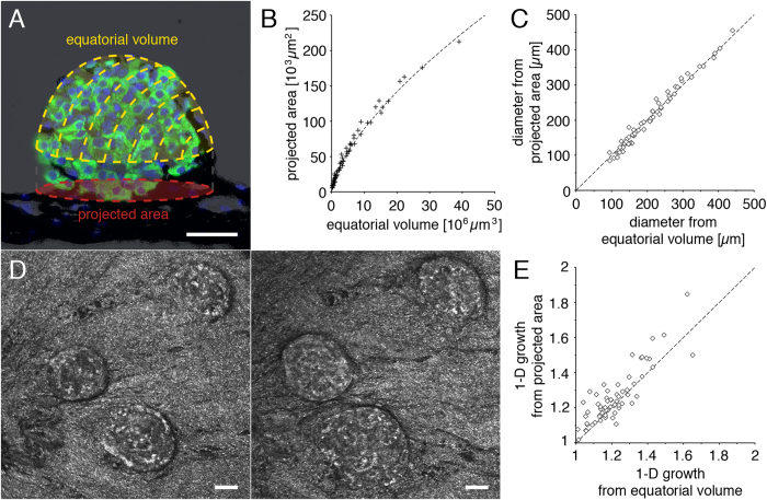
Figure 3. Quantification and characterization of islet growth in vivo by backscatter imaging.
(A) A schematic representation of dimensional information obtained by in vivo islet backscatter imaging is overlaid on an islet graft section stained by immunohistochemistry (green = insulin; blue = DAPI; black = pigmented iris). In vivo imaging permits longitudinal analysis of the islet equatorial volume and projected area. (B) Equatorial volume and projected area from individual islets imaged in vivo and (C) calculated diameter based on a spherical model. (D) In vivo imaging of the same islets engrafted into the anterior chamber of the eye at two different time points allows the longitudinal assessment of islet growth in the ob/ob mouse. (E) Growth of 27 islets was assessed over a period from 1 to 3 months in 7 ob/ob mice. The average X-, Y-, or Z-growth based either on the equatorial volume or on the projected area shows no preference for directional growth in the ob/ob mouse model. Size bars = 50 μm.

