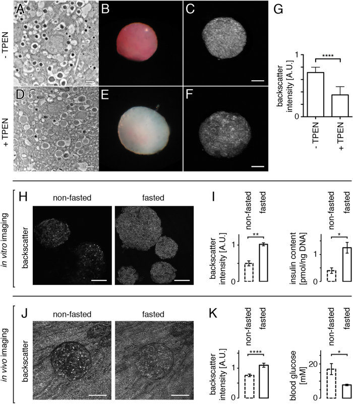
Figure 4. Backscatter intensity in beta cells originates from dense core secretory granules and provides information on islet secretory status.
(A) Electron microscopy imaging shows that untreated isolated pancreatic islets display typical dense core secretory granules containing zinc-insulin crystals. (D) The chelation of Zn2+ by treatment with TPEN impedes with normal crystallization, resulting in pale granules. (B, E) Dithizone staining in treated versus non-treated islets demonstrates a clear loss of Zn crystals after TPEN treatment. (C, F, G) The reduction in the number of dense core granules results in a decreased backscatter signal intensity. (H) Islets were isolated from mice having either free access to food or been food-deprived for 12 h, and their backscatter signal was acquired by confocal microscopy. (I) Both backscatter signal intensity and insulin content were increased when mice were fasted overnight. (J) In vivo imaging of an islet engrafted into the anterior chamber of the ob/ob mouse eye on two consecutive days shows differences in backscatter signal intensities depending on whether the mouse had free access to food or had been food-deprived for 12 h prior to imaging. Note that the backscatter signal originating from the strongly reflective pigmented iris is similar in both images, illustrating in this case that under identical imaging conditions the variations observed in signal intensities are islet-specific. (K) Quantification of backscatter signal intensities in vivo shows an increase under fasting conditions, correlating with lower blood glucose levels resulting in decreased insulin secretion from islets (n = 19 islets in 3 mice). Values are average ± SEM. *P < 0.05; **P < 0.01; ****P < 0.0001. Size bars = 100 μm.

