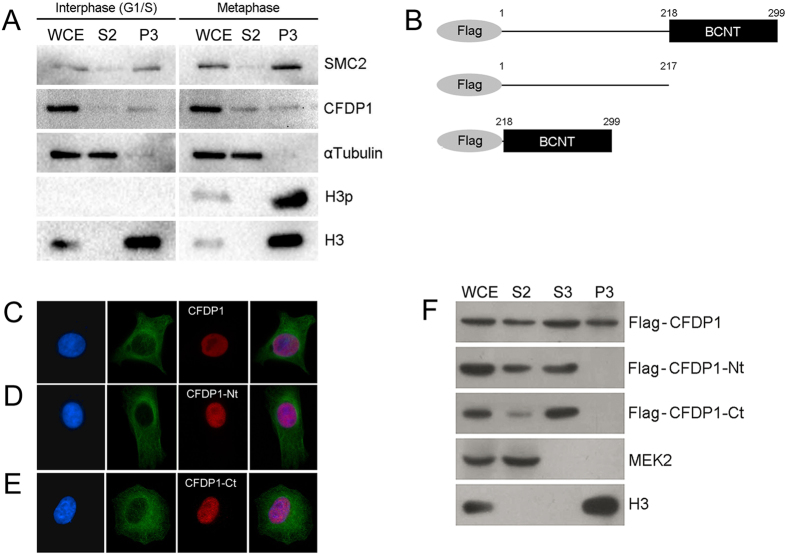
Figure 2.
Chromatin association of CFDP1 in HeLa cells (A) Western blot analysis of fractionated HeLa cells after synchronization in interphase (G1/S) or metaphase (see material and methods). As metaphase synchronization controls, the histone 3 phosphorylated in serine 10 (H3p) and SMC2 condensin have been used. H3p is a DNA condensation marker which is found only in mitotic chromatin, while SMC2 increases in condensed chromatin. As expected, H3p is specifically present in the chromatin-bound fraction (P3) of HeLa cells synchronized in metaphase. In the same fraction, the amount of SMC2 is higher compared to that of cells synchronized in G1/S. CFDP1 is detected at comparable amount in the chromatin-bound fractions (P3) of both interphase and metaphase, similar to histone H3. (B) Schematic representation of Flag-CFDP1 tagged proteins; Flag-CFDP1 full-length, isoform 1(aa 1-299); Flag-CFDP1-Nt, isoform 2 (1–217) and Flag-CFDP1-Ct, BCNT domain (218–299). (C) From left to right panels: DAPI (blue), α-tubulin (green), Flag-CFDP1 (red) and merge. (D) From left to right panels: DAPI (blue), α-tubulin (green), Flag-CFDP1-Nt (red) and merge. (E) From left to right panels: DAPI (blue), α-tubulin (green), Flag-CFDP1-Ct (red) and merge. (F) Western blot analysis of fractionated HeLa cells after transfection with Flag-CFDP1 or truncated variants. Flag-CFDP1 is detected in soluble (S2 and S3) and chromatin-bound (P3) fractions, while Flag-CFDP1-Nt and Flag-CFDP1-Ct are found only in the soluble fractions (S2 and S3). MEK2 and H3 are specific controls for soluble and chromatin fractions, respectively. Anti-MEK2, mitogen-activated protein (MAP)/ERK kinase 2, was used as cytoplasmic contamination control in P3 fraction; anti-histone H3 was used to monitor chromatin contamination in soluble fraction. WCE = whole cell extract; S2 = cytoplasmic soluble fraction; S3 = nuclear soluble fraction, P3 = chromatin-bound fraction.

