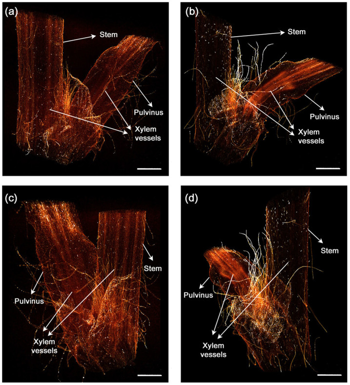
Figure 2. 3D flexible structures of the straight and bending pulvinus.
(a–d) X-ray tomograms show the internal morphological structure of the two different pulvini. 3D reconstructed images of the straight (a, c) and the bending (b, d) pulvini. The pulvinus reconstructed in a flank view (a, b) and a 130° rotated view around the vertical axis (c, d). The xylem vessels inside the pulvinus are straight or bent depending on the morphological condition. Scale bar, 2 mm.

