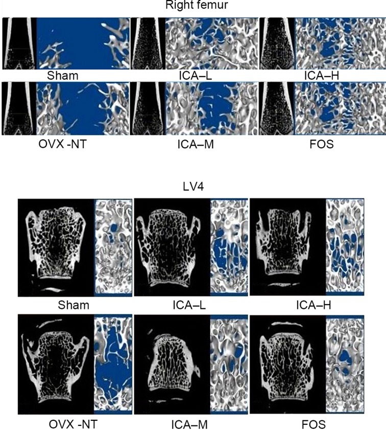
Figure 3.
ICA treatment improves bone trabeculae. Microarchitecture of trabecular bone was analyzed using micro computed tomography. Bone trabecular number in the right distal femora and LV4 increased, and the degree of bone trabecular separation became smaller in the sham-operated group. Bone trabeculae were rod-shaped, thinner, and fractured and bone trabecular separation increased in the OVX-NT group. Compared with the OVX-NT group, bone trabecular number increased in the ICA-L, ICA-M, ICA-H and FOS groups, particularly in the ICA-M and FOS groups, where bone trabecular thickness tended towards that of the sham-operated group. Although bone trabecular separation was reduced, some trabecular bone was missing; bone trabecular separation increased in the ICA-L group, but there was some improvement compared with the OVX-NT group. OVX, ovariectomized group; OVX-NT, OVX-no treatment group; ICA-L, low-dose icariin group; ICA-M, medium-dose icariin group; ICA-H, high-dose icarrin group; FOS, Fosamax-treated positive control group; LV4, fourth lumar vertebra.

