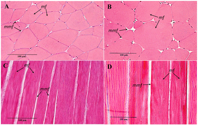
Figure 2. Microstructure observation of grass carp muscle (×400).
Transverse section microstructure of crisp grass carp (A). Transverse section microstructure of ordinary grass carp (B). Longitudinal section microstructure of crisp grass carp (C). Longitudinal section microstructure of ordinary grass carp (D). Haematoxylin and eosin stainings was used, and microstructure observations were by light microscopy, mf, muscle fibre; mmf, matrix between muscle fibres.

