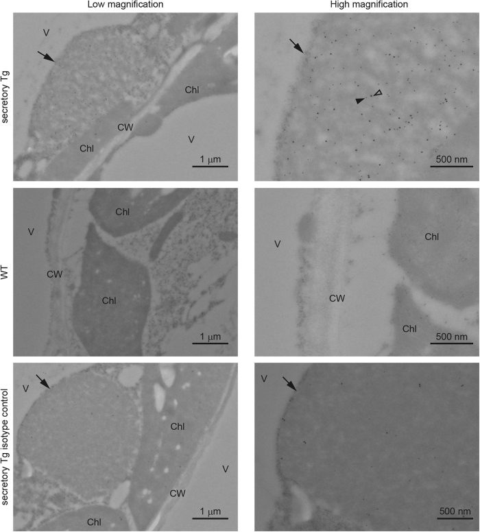
Figure 3. Immuno-electron microscopic analysis of leaf tissue of transgenic A. thaliana.
Transverse sections of A. thaliana leaf tissue were stained with anti-α chain (18-nm gold label, open arrowhead) or anti-pIgR (12-nm gold label, closed arrowhead) antibodies. Arrows indicate intracellular protein body-like structures containing S-hyIgA. V, vacuole; Chl, chloroplast; CW, cell wall. Bars represent 1 μm (low magnification) or 500 nm (high magnification).

