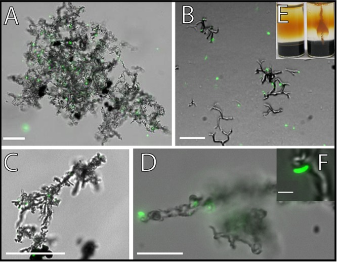
FIG 2.

Overlay of fluorescence and transmission light microscopic pictures of the microaerophilic Fe(II) oxidizers that were isolated in this study (A to D and F) and a picture of a culture growing in a gradient tube (E). Cells were stained with the LIVE/DEAD stain. Only the green fluorescence as seen with the filter set L5 is shown. (A, B, and F) Culture that was isolated from Norsminde Fjord, grown on ZVI plates (A) and in gradient tubes (B). (C and D) Culture from Kalø Vig, both grown on ZVI plates. Scale bars: 25 μm (A to C), 10 μm (D), and 1 μm (F). (E) Uninoculated gradient tube on the left and on the right a gradient tube that was inoculated with the culture from Norsminde Fjord. Panels A and F reprinted from reference 25.
