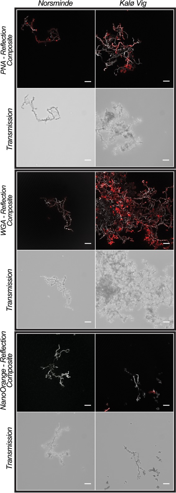
FIG 7.

Confocal laser scanning microscopy images of stalks of the two microaerophilic Fe(II) oxidizers that were isolated from Norsminde and Kalø Vig stained with three fluorescent dyes: PNA-Alexa Fluor conjugate (peanut agglutinin, specific for terminal β-galactose residues), WGA-Alexa Fluor conjugate (wheat germ agglutinin, specific for N-acetylglucosamine and N-acetylneuraminic acid), and NanoOrange (specific for proteins). In colored composite images, the stalks are visualized in gray by their reflection signal of the laser, and fluorescence stain signals are presented in red. For each sample, an additional nonconfocal transmitted light image of the stalks is shown. In composite images of samples stained with NanoOrange, the histogram was stretched linearly to a saturation of 0.3% of the pixels for better visibility of the stalks' reflection. Scale bars = 10 μm.
