Fig. 1.

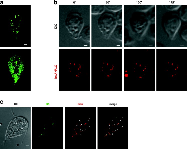
Mitosomes are stable organelles during interphase. a G. intestinalis trophozoites were fixed and immunolabeled with an anti-GL50803_9296 antibody. While the upper image shows a single G. intestinalis cell, the lower image represents the superposition of 25 imaged cells and shows areas of frequent and scarce mitosomal localization. b G. intestinalis cells expressing IscU-Halo were stained with the TMR Halo ligand and observed in medium containing 2% agarose under a confocal microscope equipped with a spinning disc. Still images (maximal projections of Z-stacks) from a time-lapse movie are shown with times indicated. Corresponding differential interference contrast (DIC) images are shown. Note that the number and distribution of organelles does not change during the indicated period of time. Scale bars, 2 μm. c G. intestinalis cells expressing human influenza hemagglutinin (HA)-tagged IscU were fixed and immunolabeled with an anti-GL50803_9296 antibody and anti-HA antibody. The arrowheads indicate mitosomes lacking the recombinant protein
