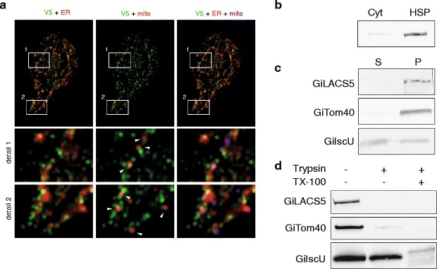
Fig. 6.

GiLACS4 populates the endoplasmic reticulum (ER)–mitosome contact sites. a G. intestinalis cells expressing V5-tagged GiLACS were fixed and immunolabeled using anti-V5 tag, anti-GL50803_9296, and anti-PDI2 antibodies. Left: V5 in green and PDI2 in red; Middle: V5 in green and GL50803_9296 in red; Right: V5 in green, PDI2 in red and GL50803_9296 in magenta. The cells were observed by structured illumination microscopy (SIM). The arrows indicate spots where the mitosomal signal meets the V5-tagged GiLACS4. b The cells were fractionated and the high-speed pellet (HSP) and cytosolic fraction were immunolabeled with anti-V5 antibody. c The HSP fraction was subjected to sodium carbonate extraction and the resulting fractions immunolabeled with anti-V5 (GiLACS4), anti-IscU, and anti-Tom40 antibodies. S - soluble fraction, P - membrane bound fraction. d The HSP fraction was treated with trypsin with or without the presence of 1% Triton. The samples were immunolabeled with anti-V5 (GiLACS4), anti-IscU, and anti-Tom40 antibodies
